The Invisible Light: How X-rays Went from Accidental Discovery to World-Changing Technology
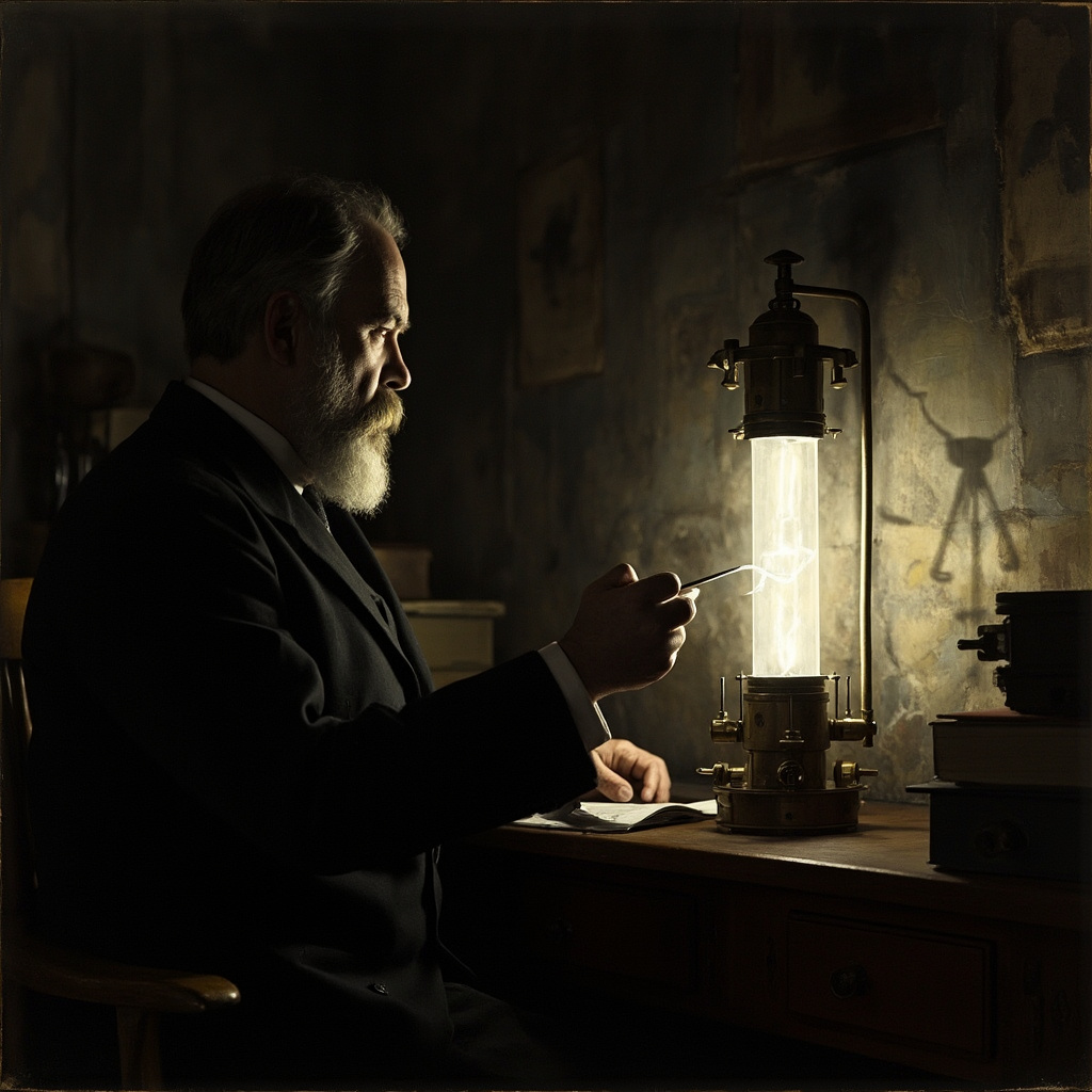
On a chilly November evening in 1895, a 50-year-old German physicist named Wilhelm Conrad Röntgen was working alone in his darkened laboratory at the University of Würzburg. He was experimenting with cathode ray tubes — the cutting-edge technology of his day — when something peculiar caught his eye. A fluorescent screen on the other side of the room was glowing. It shouldn’t have been. The tube was completely covered in thick black cardboard. Whatever was causing that glow was passing straight through the covering like it wasn’t even there.
Röntgen was baffled. He spent the next several weeks barely eating or sleeping, locked in his laboratory, obsessively investigating this mysterious new radiation. He didn’t know what it was, so he called it “X-rays” — X for unknown. It was a placeholder name that stuck forever. What he discovered in those feverish weeks would transform medicine, reshape warfare, revolutionize industry, and accidentally kill quite a few people along the way.
The Photograph That Stunned the World
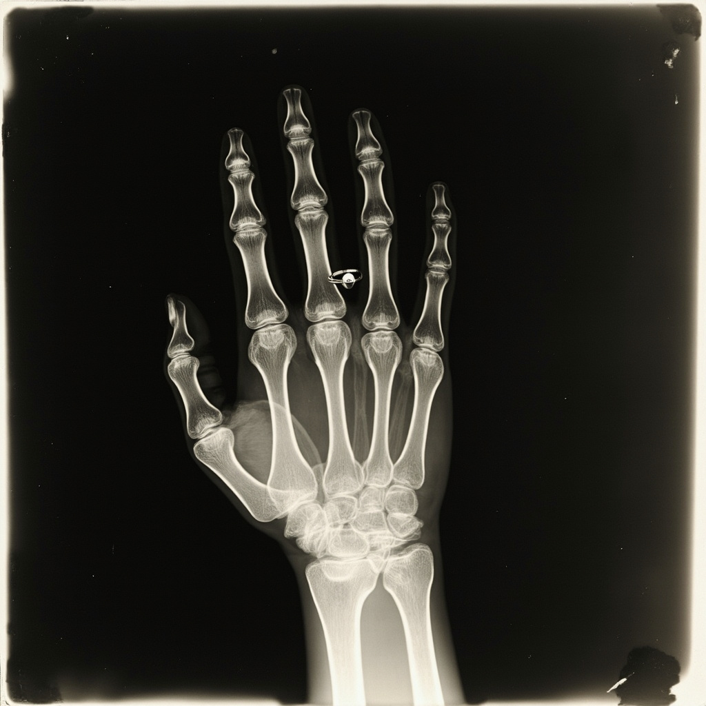
On December 22, 1895, Röntgen asked his wife Anna Bertha to place her hand on a photographic plate while he aimed his X-ray tube at it. The exposure took about 15 minutes — during which Anna Bertha had to hold perfectly still. The resulting image was haunting: the dark shadows of her bones clearly visible, her wedding ring floating ghostlike around her finger. When she saw the image, she reportedly gasped, “I have seen my death.”
Röntgen published his findings on December 28, 1895, and the news exploded across the globe with a speed that rivaled the telegraph itself. Within days, newspapers on every continent were breathlessly reporting on the “new photography” that could see through flesh to the bones beneath. The public was equal parts fascinated and terrified.
In 1901, Röntgen was awarded the very first Nobel Prize in Physics. True to his modest character, he donated the prize money to his university and refused to patent his discovery, believing it belonged to humanity. He died in relative obscurity in 1923, during the economic chaos of Weimar Germany, while the technology he unleashed was already changing the world in ways he never imagined.
X-ray Mania: When Seeing Bones Was Entertainment

The years following Röntgen’s discovery saw an extraordinary craze sweep through Europe and America. “X-ray parlors” sprang up in cities, offering curious customers the chance to see their own skeletons for a small fee. It was the Victorian equivalent of a selfie — except instead of your face, you were showing off your metacarpals.
Department stores installed X-ray machines as novelty attractions. Shoe stores offered “fluoroscopes” that let customers wiggle their toes inside their shoes to check the fit — bombarding their feet with radiation in the name of retail satisfaction. These shoe-fitting fluoroscopes remained in widespread use from the 1920s through the 1950s before someone finally asked, “Wait, is this a good idea?”
The entertainment industry embraced X-rays with gusto. Thomas Edison, ever the showman, demonstrated a large fluoroscope at the 1896 Electrical Exhibition in New York City. His assistant, Clarence Dally, operated the device extensively. Dally would become one of the first known casualties of radiation exposure in America — he developed severe radiation burns, had both arms amputated, and died in 1904 at the age of 39. Edison, shaken by Dally’s fate, abandoned all X-ray research.
The early enthusiasm was dangerously naive. Without understanding radiation’s biological effects, people treated X-rays as harmless curiosities. Doctors would demonstrate X-ray machines at dinner parties. Inventors proposed X-ray opera glasses so theatergoers could peer through walls. A London company advertised “X-ray proof undergarments” for modest ladies who feared their privacy was at risk. The fear was absurd, but the entrepreneurial spirit was very real.
From Parlor Trick to Battlefield Savior

While civilians were gawking at their bones in parlors, the medical community quickly recognized X-rays’ life-saving potential. During the Balkan Wars of 1897 and the Boer War (1899–1902), military surgeons used X-rays to locate bullets and shrapnel lodged in wounded soldiers — a task that had previously required agonizing exploratory surgery.
But it was World War I that truly proved X-rays’ value on a massive scale. Marie Curie — already famous for her research on radioactivity — threw herself into the war effort with characteristic determination. She equipped a fleet of vehicles with portable X-ray machines, creating the world’s first mobile radiological units. Soldiers affectionately called them “petites Curies” — little Curies.
Curie personally drove these vehicles to field hospitals near the front lines, training doctors and technicians to use the equipment. Her teenage daughter Irène joined her, operating X-ray machines in battlefield hospitals at the age of 17. Together, the Curies helped perform over a million X-ray examinations during the war, guiding surgeons to extract bullets and shrapnel that would have otherwise meant amputation or death.
The wartime experience transformed X-ray technology from a medical curiosity into an indispensable clinical tool. After the war, hospitals worldwide invested in permanent X-ray departments, and the specialty of radiology was born.
The Dark Side: Radiation’s Hidden Toll
The enthusiasm for X-rays came at a terrible price. In the early decades, no one understood the cumulative damage that radiation inflicted on human tissue. Radiologists routinely tested their equipment by X-raying their own hands. Many developed radiation dermatitis — chronic skin damage that progressed to ulceration and cancer.
By the 1930s, the toll was becoming undeniable. Hundreds of early radiologists and X-ray technicians had developed cancers, lost fingers and limbs, or died from radiation-related illnesses. A memorial erected in Hamburg, Germany, in 1936 listed 169 names of radiologists who died from radiation exposure. By 1960, the list had grown to 360 names.
The radium industry — a cousin of X-ray technology — was claiming victims too. The infamous “Radium Girls” of the 1920s, young women who painted luminous watch dials with radium-laced paint, developed devastating jaw necrosis and cancers after being told to lick their brushes to form a fine point. Their legal battle against the U.S. Radium Corporation became a landmark case in occupational health law.
These tragedies eventually forced the development of radiation safety standards. Lead shielding, exposure limits, dosimetry badges, and the principle of using the minimum radiation dose necessary all emerged from the painful lessons of the early X-ray era.
The Modern Age: From Film to Digital

The second half of the 20th century brought revolutionary advances. In 1971, British engineer Godfrey Hounsfield introduced the CT (computed tomography) scanner, which used X-rays and computer processing to create detailed cross-sectional images of the body. It was like going from a shadow puppet show to a full 3D movie. Hounsfield shared the 1979 Nobel Prize in Physiology or Medicine for this invention.
CT scanning transformed diagnostic medicine. Doctors could now see tumors, blood clots, fractures, and internal bleeding with unprecedented clarity — without surgery. Emergency rooms became dependent on CT scanners for evaluating trauma patients. Oncologists used them to stage cancers and monitor treatment response.
The digital revolution of the 1980s and 1990s replaced photographic film with electronic sensors, bringing instant image display, computer enhancement, and electronic storage. Radiation doses plummeted as detector technology improved. Today’s digital X-ray systems deliver a fraction of the radiation that early machines produced.
The 21st century has brought further marvels: cone beam CT for three-dimensional imaging, AI algorithms that can detect diseases on X-rays with superhuman accuracy, and portable X-ray devices small enough to fit in a backpack for use in disaster zones and remote communities.
A Legacy of Light and Shadow
The story of X-rays is, in many ways, a perfect parable of human discovery. A curious scientist stumbles upon something extraordinary. Society embraces it with reckless enthusiasm. People suffer from the unintended consequences. And gradually, painfully, we learn to harness the discovery safely and effectively.
From Röntgen’s darkened laboratory to modern hospital radiology suites, from Victorian bone-gazing parlors to AI-powered diagnostic systems, the invisible light that one physicist discovered by accident has illuminated the hidden interior of the human body for 130 years. It has saved millions of lives, enabled entire medical specialties, and — in its darkest chapters — reminded us that every powerful technology demands respect.
Wilhelm Röntgen never sought fame or fortune from his discovery. He gave it freely to the world, asking nothing in return. The X stands for unknown — and in a sense, it still does. Even today, researchers are finding new applications for X-ray technology, from archaeology to art conservation to airport security. The unknown ray turned out to be one of the most versatile and consequential discoveries in human history.
Not bad for an accident on a November evening.